Assalamualaikum & hi readers!! Hope all of you having a great day today :)
Okey, today I will write on something that were major important to us, human. Which is cardiovascular system. So, lets proceed !
Coordinated cycles of heart contraction drive double circulation in mammals.
The time delivery of O2 to the body's organs is critical: Some brain cells, for example, die if their O2 supply is interrupted for as little as a few minutes. How does the mammalian cardiovascular system meet the body's continuous demand for O2 ??! To answer this question, we must consider how the parts of the system are arranged and how each part functions.
Mammalian Circulation.
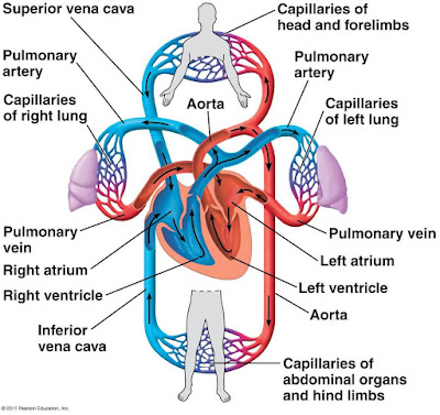
Lets first examine the overall organization of the mammalian cardiovascular system, beginning with the pulmonary circuit. Contraction of the right ventricle pumps blood to the lungs via the pulmonary arteries. As the blood flows through capillary beds in the left and right lungs, it loads O2 and unloads CO2. Oxygen rich blood returns from the lungs via the pulmonary veins to the left atrium of the heart. Next, the oxygen rich blood flows into the heart's left ventricle, which pumps the oxygen rich blood out to body tissues through the systematic circuit. Blood leaves the left ventricle via the aorta, which conveys blood to arteries throughout the body. The first branches leading from the aorta are the coronary arteries, which supply blood to the heart muscle itself. Then branches lead to capillary beds in the head and arms. The aorta then descends into the abdomen, supplying oxygen rich blood to arteries leading to capillary beds in the abdominal organs and legs. Within the capillaries, there is a net diffusion of O2, from the blood to the tissues and of CO2. into the blood. Capillaries rejoin, forming venules, which conveys blood to veins. Oxygen poor blood from the head neck and forelimb is channeled into a large vein, the superior vena cava. Another large vein, the inferior vena cava, drains blood from the trunk and hind limbs. The two venae cavae empty their blood into the right atrium, from which the oxygen poor blood flows into the right ventricle.
Mammalian Heart.
Lets now take a closer look at how heart works. Located behind the sternum (breastbone) the human heart is about the size of a clenched fist and consist mostly of cardiac muscle. The two atria have relatively thin walls and serve as collection chamber for blood returning to the heart from the lungs or other body tissues. Much of the blood that enters the atria flows into the ventricles while all heart chambers are relaxed. The remainder is transferred by contraction of the atria before the ventricles begin to contract. Compared to the atria, the ventricles have thicker walls and contract much more forcefully especially the left ventricle which pumps blood throughout the body via the systemic circuit. Although the left ventricle contracts with greater force than the right ventricle, it pumps the same volume of blood as the right ventricle during each contraction.
The hearts contracts and relaxes in a rhythmic cycle. When it contracts, it pumps blood, when it relaxes, its chambers fill with the blood. One complete sequence of pumping and filling is referred to as the cardiac cycle. The contraction phase of cycle called systole, and the relaxation phase called diastole.
 |
| Cardiac Cycle |
 |
| Systole & Diastole phase. |
The volume of blood each ventricle pumps per minute is the cardiac output. Two factors determine cardiac output: the rate of contraction or heart rate , and the stroke volume, the amount of blood pumped by a ventricle in a single contraction. The average stroke volume in humans is about 70mL. Multiplying this stroke volume by a resting heart rate of 72 beats per minute yields a cardiac output of 5mL/min about equal to the total volume of blood in the human body. During heavy exercise, the cardiac output increases as much as fivefold.
Four valves in the heart prevent backflow and keep blood moving in the correct direction. Made of flaps of connective tissues, the valves open when pushed from one side and close when pushed from the other. An atrioventricular (AV) valve lies between each atrium and ventricle. The AV valves are anchored by strong fibers that prevent them from turning inside out. Pressure generated by the powerful contraction of the ventricles closes the AV valves, keeping blood from flowing back into the atria.
Semilunar valves are located at the two exits of the heart where the aorta leaves the left ventricle and where the pulmonary artery leaves the right ventricle. These valves are pushed open by the pressure generated during contraction of the ventricles. When the ventricles relax, blood pressure built up in the aorta and the pulmonary artery closes the semilunar valves and prevents significant backflow.
 |
| Control Of Heart Rhythm. |


No comments:
Post a Comment